Cryo-EM methods
The cryo-EM method has become very powerful in the past several years and for many structural biologists it is becoming the method of choice. The full potential of the technique, however, is still to be realized as the scientific community is in great need of hardware-based and software improvements. Although the first 3D images of biological specimens at true atomic resolution have been obtained recently, the routine resolution of cryo-EM still lags behind in detail and quality in comparison with common material science electron microscopy as it is imaged at the Ernst-Ruska Centre (ER-C) 1 and 2. Therefore, in collaboration with the ER-C-1 and ER-C-2 we will apply new imaging hardware to biological samples. Our technique development also includes novel sample preparation, data acquisition and image processing methods that we benchmark using biological test specimens. We apply these innovative cryo-EM methods to the structures of challenging biological systems with a particular focus on membrane assemblies.
Related Publications
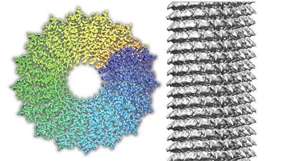
- Weis F., Beckers M., von der Hocht I., Sachse C. (2019) Elucidation of viral disassembly switch of tobacco mosaic virus. EMBO Reports 20(11):e48451.
- Fromm, S.A., Bharat, T.A.M., Jakobi, A.J., Hagen, W.J.H., and Sachse, C. (2015). Seeing tobacco mosaic virus through direct electron detectors. J Struct Biol 189, 87–97.